Tuftsin 5mg
Endogenous IgG-derived tetrapeptide studied for macrophage modulation, neuropilin-1 signaling, and neuroinflammatory regulation.
Mechanism
Tuftsin binds neuropilin-1 and signals via the TGF-beta receptor-1/Smad3 pathway to promote regulatory T-cell expansion and bias macrophages and microglia toward an anti-inflammatory M2 phenotype.
Dosing
Published experimental reference points begin at an analytical benchmark of 100 mcg (0.04 mL or 4 U-100 units at 2.5 mg/mL).
Reconstitution
Reconstituting a 5 mg vial with 2.0 mL of bacteriostatic water yields a stock concentration of 2.5 mg/mL.
Storage
Store lyophilized vials at 2–8°C protected from light, and keep reconstituted solutions refrigerated or aliquoted at -20°C to minimize degradation into antagonistic fragments.
Mix & measure Tuftsin 5mg
Pre-filled with this protocol's recommended BAC water and documented starting dose — edit any field to run your own numbers.
Mix & measure Tuftsin 5mg
Vial strength → BAC water → target dose
Total doses in vial: 50.0
U-100 syringe: 100 units = 1 mL
Reconstitution math only — not dosing advice. U-100 syringe: 100 units = 1 mL. Advanced calculator →
Dosing Chart
| Phase / Day(s) | Dose & Frequency | Volume (U-100 units / mL) |
|---|---|---|
| Reference Tier 1 | 100 mcg laboratory reference | 4 units (0.04 mL) |
| Reference Tier 2 | 250 mcg laboratory reference | 10 units (0.10 mL) |
| Reference Tier 3 | 500 mcg laboratory reference | 20 units (0.20 mL) |
| Reference Tier 4 | 1 mg laboratory reference | 40 units (0.40 mL) |
Reconstitution Steps
- 1
Sterilize the rubber stoppers of both the tuftsin vial and the bacteriostatic water container with individual alcohol pads and allow to dry.
- 2
Draw exactly 2.0 mL of bacteriostatic water using a sterile syringe.
- 3
Introduce the diluent slowly into the peptide vial, aiming the stream along the interior glass wall rather than directly at the lyophilized powder.
- 4
Allow the mixture to rest for 30 seconds, then roll or swirl the vial gently until completely clear without shaking.
- 5
Place the reconstituted solution into refrigeration at 2–8°C away from light exposure.
Supplies Needed


Peptide Vial
Lyophilized Tuftsin (5 mg) research reagent.


Insulin Syringes (U-100)
Precision volumetric sampling (100 units = 1.0 mL).


Bacteriostatic Water
2.0 mL used as diluent for reconstitution.


Alcohol Swabs
Surface decontamination of vial tops before needle entry.
Why researchers study it
Immune modulation and macrophage polarization assays
Parent structure comparative analysis against Selank
Neuroinflammatory models of demyelination and EAE
These describe what is being studied, not proven benefits, approved uses, or promised results.
Overview
Tuftsin is an endogenous tetrapeptide corresponding to the amino acid sequence Thr-Lys-Pro-Arg, originally identified within the CH2 domain of the immunoglobulin G heavy chain. In native physiology, it is cleaved enzymatically from IgG to exert specific regulatory effects on phagocytic cells. Despite being characterized several decades ago, tuftsin remains strictly an analytical-grade laboratory reagent without clinical approval or established human dosing parameters. Modern investigation centers heavily on neuroimmunology and macrophage polarity rather than simple immune stimulation. While early assays highlighted its capacity to enhance phagocytosis in isolated granulocytes, current molecular findings demonstrate that tuftsin binds neuropilin-1 to trigger downstream TGF-beta receptor signaling, shifting microglial and macrophage populations toward a protective, anti-inflammatory M2 phenotype. Due to its susceptibility to rapid enzymatic degradation by aminopeptidases, synthetic analogues like Selank were subsequently engineered. When working with native tuftsin in a laboratory setting, careful handling and precise reconstitution are required to preserve peptide integrity and avoid the accumulation of antagonistic breakdown fragments.
References
- 1.Fridkin & Najjar, Critical Reviews in Biochemistry and Molecular Biology, 1989 — Tuftsin: its chemistry, biology, and clinical potential
- 2.Nissen, Selwood & Tsirka, Journal of Neurochemistry, 2013 — Tuftsin signals through its receptor neuropilin-1 via the TGF-beta pathway
- 3.Wu, Nissen, Chen & Tsirka, PLoS ONE, 2012 — Tuftsin promotes an anti-inflammatory switch and attenuates symptoms in EAE
- 4.Nissen & Tsirka, Glia, 2016 — Tuftsin-driven EAE recovery requires neuropilin-1
- 5.Thompson, Nissen, Pretory & Tsirka, Frontiers in Immunology, 2018 — Tuftsin combines with remyelinating therapy in CNS demyelinating disease
- 6.Bump & Najjar, Molecular and Cellular Biochemistry, 1984 — Tuftsin, a natural modulator of macrophage activity
- 7.Nishioka, Wagle, Rodriguez, Maeta, Kubo & Dessens, Journal of Surgical Research, 1994 — Studies of human granulocyte phagocytosis stimulation by tuftsin
- 8.Kubo, Roh, Oyedeji, Romsdahl & Nishioka, Hepato-Gastroenterology, 1998 — Effect of tuftsin on human Kupffer cell
- 9.Mathé, Cancer Detection and Prevention Supplement, 1987 — Do tuftsin and bestatin constitute a biopharmacological immunoregulatory system?
- 10.Xu, Zhu, Xiang, Li, Sun, Ma, Sun & Liu, Biomaterials, 2011 — Synthesis and immunomodulatory activity of [60]fullerene-tuftsin conjugates
- 11.Czabak-Garbacz, Cygan, Wolański & Kozlovsky, Pharmacological Reports, 2006 — Long-term treatment with the tuftsin analogue TP-7 and anxiety-phobic states
- 12.Patel, Williams, Shmigel & Hinshaw, Surgery, 1981 — Preservation of splenic function by autotransplantation of traumatized spleen in man
Supplies Needed
Suggested supplier
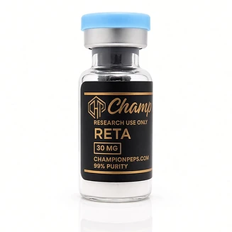
Research use only. Listing a supplier is not an endorsement of any protocol on this site, and nothing sold there is approved for human use.